
Calcific tendonitis of the shoulder
Calcific tendonitis is a painful shoulder condition caused by the deposit of calcium crystals within the rotator cuff tendons, leading to severe localized inflammation and pressure.
Calcific tendonitis overview
The rotator cuff is a group of muscles and tendons that hold the upper arm bone securely in the shoulder socket while enabling overhead movement and rotation. Calcific tendonitis occurs when calcium deposits build up within one or more of these rotator cuff tendons (most commonly the supraspinatus tendon). As the calcium deposit expands, it increases internal pressure within the tendon and narrows the subacromial space, the narrow gap between the rotator cuff and the highest point of the shoulder blade (acromion). This causes painful impingement, pinching, and inflammation whenever the arm is raised.
What are the symptoms?
- Sudden, severe, and disabling shoulder pain (often worst during the resorptive phase)
- Marked pain when lifting the arm overhead or rotating the shoulder
- Severe night pain, making it difficult to sleep on the affected side
- Restricted joint mobility and localized tenderness at the front or side of the shoulder
How is calcific tendonitis diagnosed?
Diagnosing calcific tendonitis begins with a detailed physical examination by your consultant to assess range of motion, pain points, and rotator cuff integrity.
Imaging studies are essential to confirm the diagnosis and locate the deposit:
- X-rays: Standard shoulder X-rays clearly reveal dense, chalky calcium deposits within the rotator cuff tendons and determine their size and location.
- Ultrasound scan: Provides high-resolution, real-time images of the tendon to confirm whether the calcium is soft or hard and check for surrounding tendon inflammation or bursitis.
How is it treated?
Management aims to relieve acute pain, remove or reduce the calcium deposit, and restore normal shoulder movement
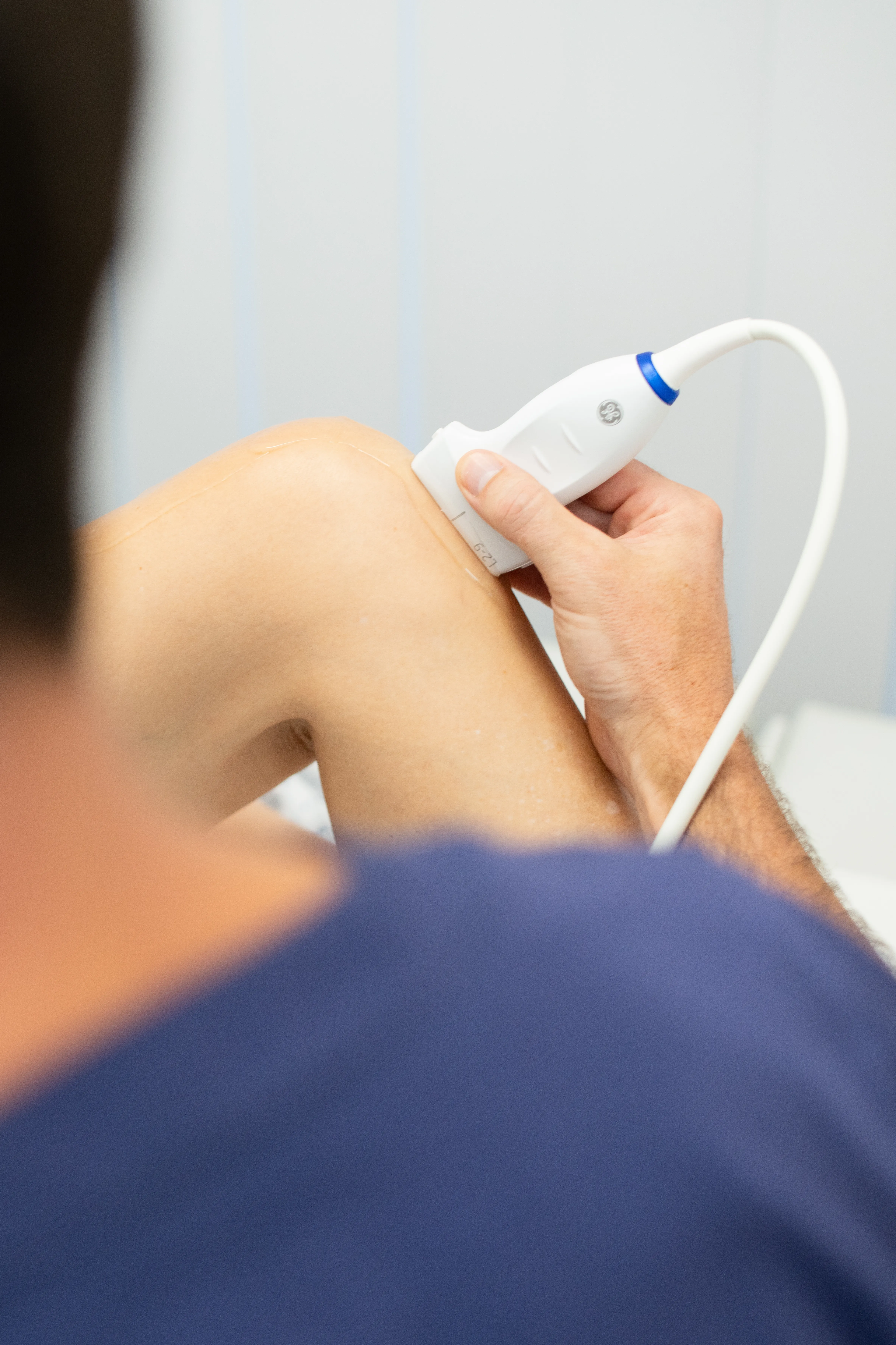
Treatment routes for Calcific tendonitis
- Medication & rest: Anti-inflammatory medications (NSAIDs) help manage acute pain and swelling during flares.
- Targeted steroid injections: Corticosteroid injections into the subacromial space provide rapid pain relief and reduce acute tissue inflammation.
- Ultrasound-guided barbotage (lavage): A highly effective, minimally invasive outpatient procedure. Under local anaesthetic and ultrasound guidance, a specialist inserts a fine needle directly into the calcium deposit, flushes it with saline, and washes out (aspirates) the softened calcium.
- Focused physical therapy: Once acute pain is controlled, a gentle stretching and strengthening program restores joint flexibility and rotator cuff function.
Recovery from calcific tendonitis
Pain control and gentle mobility
Following barbotage or keyhole surgery, focus on controlling inflammation with ice and prescribed medication. If surgery was performed, a sling may be worn for comfort for a few days. Gentle pendulum exercises and passive shoulder movements begin almost immediately. Dressings should be kept clean and dry.
Restoring range of motion
Stitches (if applicable) are typically checked or removed around 10 to 14 days. Guided physical therapy shifts toward progressive stretching exercises to regain full overhead reach and rotation.
Strengthening and full activity
Targeted rotator cuff and scapular strengthening exercises are introduced. Most patients achieve full, pain-free function and return to regular exercise and manual tasks during this phase.
Working with professionals and teams
Our shoulder specialists support professional athletes, sports teams, active individuals and people whose work places high demands on their shoulders. Working closely with radiologists, physiotherapists and the wider multidisciplinary team, they provide coordinated care from diagnosis through to rehabilitation, helping each person return safely to sport, work or everyday activities.
The same specialist expertise and collaborative approach are available to every patient, whatever your activity level or recovery goals.

Our Experts - Leaders in Shoulder Treatment







How to pay
We offer flexible options depending on how you choose to access care at Fortius. Whichever option you choose, we’ll help make the process clear and straightforward from the start.
Private medical insurance
If your care is covered by private medical insurance, we’ll usually invoice your insurer directly. Please check your policy in advance and request an authorisation code if your insurer requires one. You’ll be responsible for paying any policy excess or costs that aren’t covered by your insurance.

Paying for yourself
You don’t need private medical insurance to access care at Fortius. If you’re paying for your own treatment, our team will explain the costs clearly and provide a personalised quote before you decide whether to go ahead.
Find a clinic near you
Fortius Clinic London

Our 79,000 sq ft, seven-story, private hospital on Wigmore Street has 39 en-suite inpatient rooms, consultation and treatment rooms, and a physiotherapy suite.
The hospital has a high-spec imaging suite fully equipped with an MRI, CT scanner and X-ray machine. There are three state-of-the-art operating theatres, two anaesthetic rooms and a recovery bay.
Fortius Clinic London has two Mako robots for hip and knee joint replacements.
- 66 Wigmore Street W1U 2SB
- 0203 195 2442
- Outpatient times are Monday to Friday from 8.00am to 8.00pm.
- Sports & Exercise Medicine
- Snow Injuries Clinic
- Tendon Clinic
- MRI Scan
- Ultrasound
- X-Ray
- Physician services
- Rheumatology
- CT Scan
- Image guided injections
- Physiotherapy
- MAKO Robotic Joint Replacement
- Knee replacement revision clinic
Fortius Clinic Wimbledon
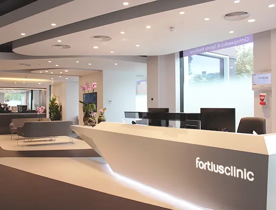
Our Wimbledon clinic houses a state-of-the-art imaging centre with a 3T MRI and X-ray scanners, Ultrasound, seven consulting rooms, two spacious waiting areas and well equipped treatment rooms all located on one level, ensuring easy accessibility for all patients.
Our partnership with Spire St Anthony’s Hospital allows us to provide orthopaedic surgery closer to our clinic. We treat a full range of conditions and perform joint replacement surgery using the latest Mako robotic assisted technology.
The building at 22 Worple Road has steps leading up to the front entrance. If you require assistance please inform us before your arrival by calling ahead on 020 3195 2442
- 22 Worple Road SW19 4DD
- 0203 195 2442
- Outpatient times are Monday to Friday from 8.00am to 8.00pm.
- Snow Injuries Clinic
- MRI Scan
- Ultrasound
- X-Ray
Fortius Clinic Marylebone
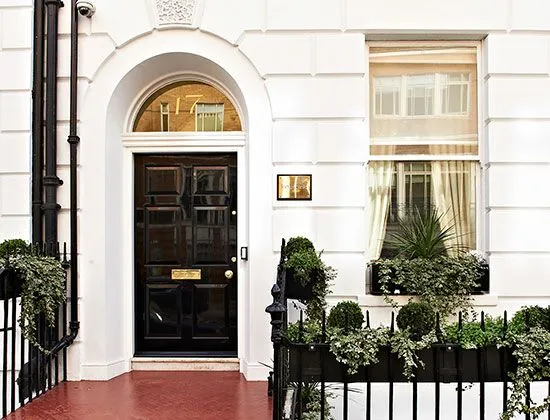
Our Marylebone Clinic is conveniently located in the historic area between Baker Street and Marylebone High Street, just off Manchester Square, home to the iconic Wallace Collection. We have seven consulting rooms, private waiting areas on each floor, well equipped treatment rooms and diagnostic imaging.
- 17 Fitzhardinge StreetW1H 6EQ
- 020 3195 2442
- Outpatient times are Monday to Friday from 8.00am to 8.00pm.
- MRI Scan
- Ultrasound
- X-Ray
- Physician services
- Rheumatology
Fortius Clinic City
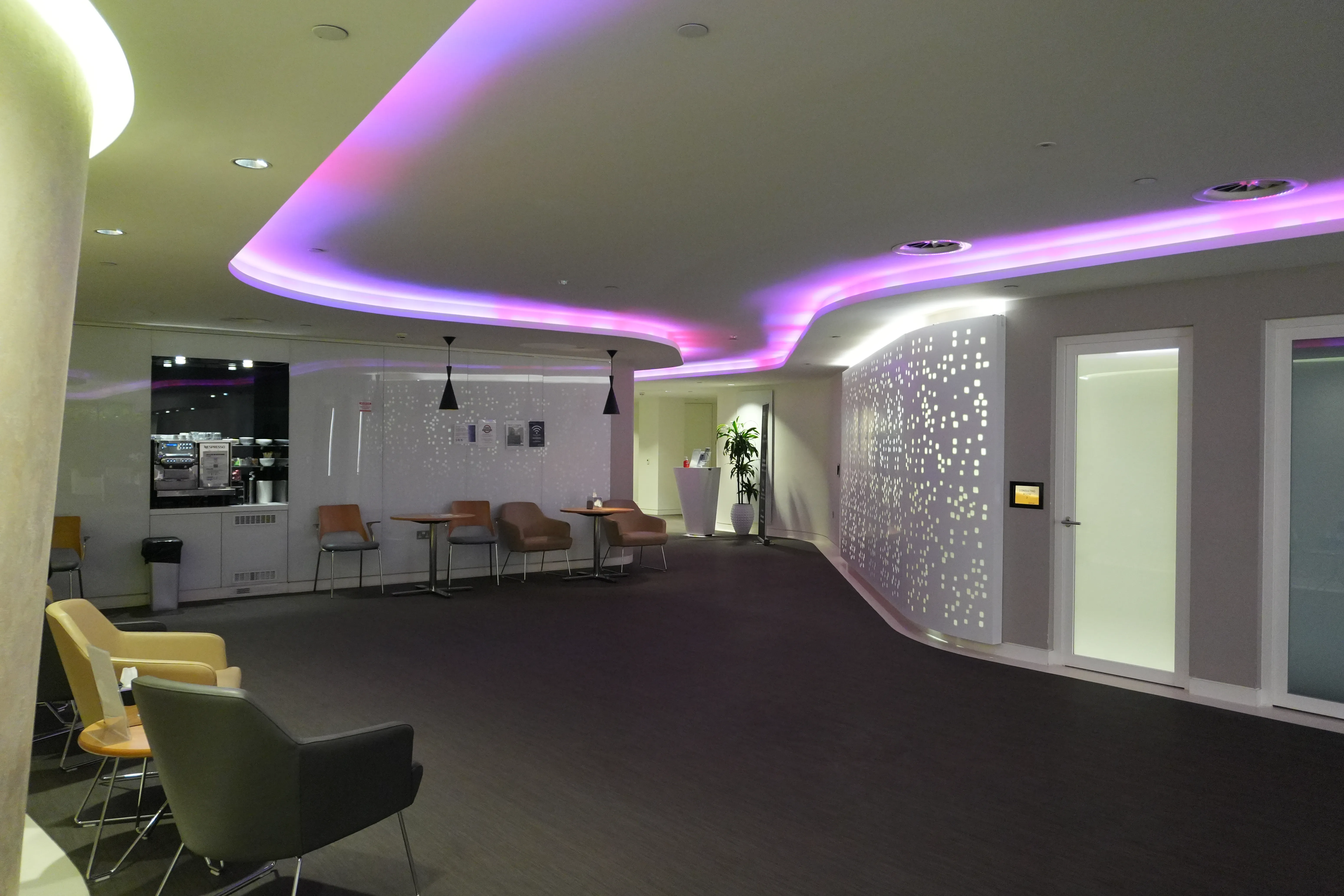
Our City Clinic is conveniently located between Bank and Monument stations, within the historic and financial heart of London. At our modern, two-floor facility we have nine consulting rooms, spacious waiting areas, well equipped treatment rooms and an imaging centre.
Explore our City Clinic with our virtual tour.
If you are travelling by Tube, our closest underground stations are Bank and Monument, exit 6 for King William Street.
Should you be travelling by car, the closest parking is a three minute drive away at NCP Car Park London Vintry Thames Exchange. If you are attending clinic on a weekday, please be aware that we are based in the congestion charge zone.
- 75 King William Street EC4N 7BE
- 0203 195 2442
- Outpatient times are Monday to Friday from 8.00am to 8.00pm.
- Snow Injuries Clinic
- Tendon Clinic
- Sports & Exercise Medicine
- MRI Scan
- Ultrasound
- X-Ray
- Physician services
- Rheumatology
1 of 4